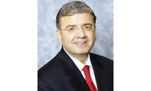
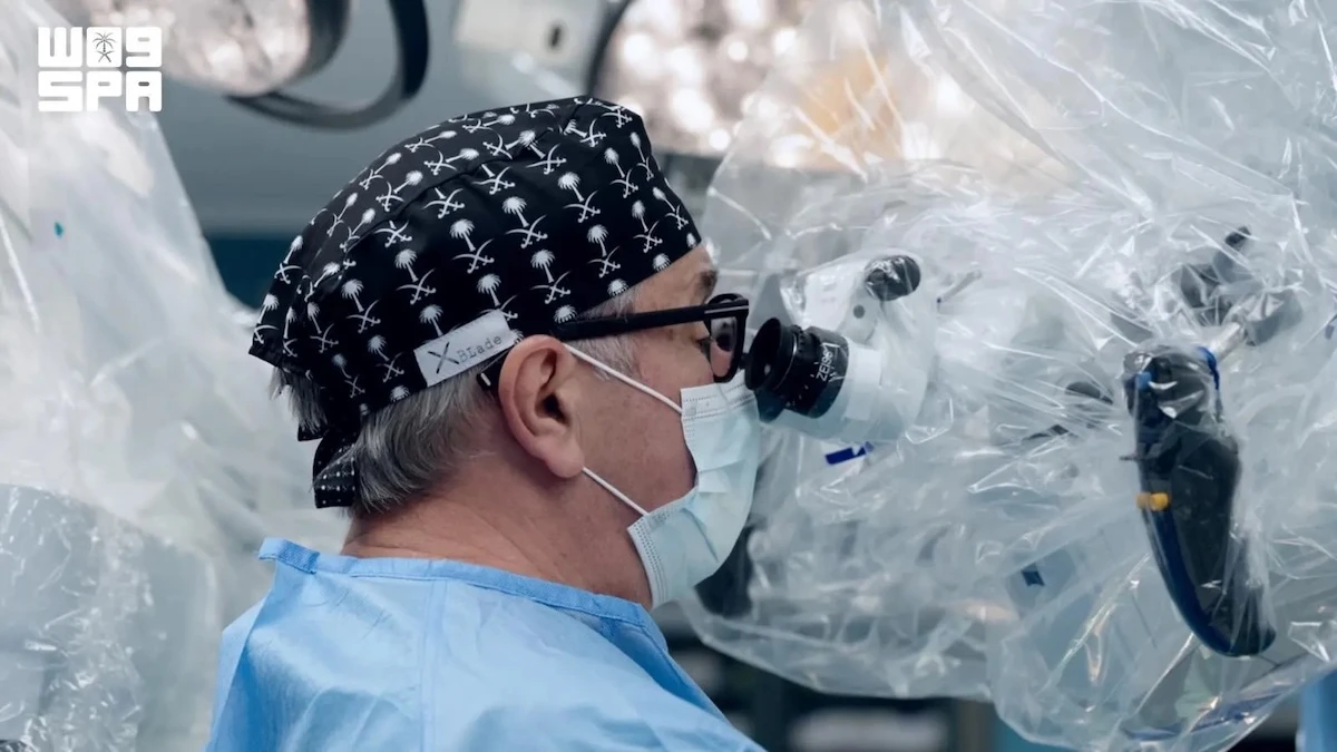
Dr. William Zoghbi, director of the Cardiovascular Imaging Institute at Houston Methodist Hospital in Texas, has recently discussed the latest insights on cardiac health, specifically Multimodality Cardiovascular Imaging, among health professionals in the Kingdom.
“Multimodality Cardiovascular Imaging has witnessed a number of developments and additions over the past five years,” he said at the 26th annual conference of the Saudi Heart Association (SHA) recently.
He said that “traditional doctors used ultrasound and nuclear imaging, two technologies which were door openers in the cardiac health field.” He added that recent modalities have enabled people to image the heart and its arteries in ways that were previously impossible.
“Cardiac CT scans can now detect the earliest manifestations of atherosclerosis as calcifications in the coronary arteries and, when combined with dye injection (CT coronary and angiography), allows us to see the coronary arteries themselves, completely non-invasively,” he said.
On the other hand, he added, a cardiac MRI can give a unique assessment of the heart muscle by localizing any damage to the heart after a heart attack or other ailments.
When asked about the risk that such modalities might have, Dr. Zogbhi said that while cardiovascular procedures are not entirely risk free, the benefits from using such modalities are very valuable and effective in a cardiac patient.
For instance, ultrasound imaging, he said, does not have any risks at all, except that patients might feel a little discomfort when the probe is put on their chest.
For nuclear imaging, the nuclear material is safe, particularly when used sporadically. Cardiac CT use X-Rays, and radiation doses have decreased significantly with the newer equipments.
“So it is generally very safe when used appropriately. MRI does not have radiation but because it uses a magnetic field, some metal implants like pacemakers may be a contraindication; the newer generation of pacemakers, however, are MRI safe,” added Dr. Zoghbi.
It is worth mentioning that Houston Methodist Hospital has a unique cardiovascular imaging Institute where all cardiovascular modalities are co-located for better patient care, education and research, along with an advanced visualization technology center (Plato’s CAVE, Computer-Augmented Virtual Environment) that allows physicians to navigate the heart non-invasively and with the highest precision.
Dr. Zoghbi is a graduate of the American University in Beirut and pursued further training in the USA. He has also authored more than 250 original publications in the field of cardiovascular imaging and has been invited to lecture nationally and internationally.
He is the past president of both the American Society of Echocardiography and American College of Cardiology.
New technologies allow precise cardiac imaging